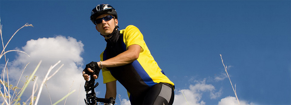
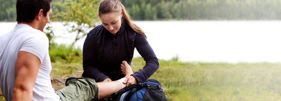


Physiotherapy in Beamsville for Injury Care
Would you like to learn more about the latest options available for surgical treatment of open tibial fractures? This article might answer some of your questions; and don't forget to make an appointment with your physiotherapist at Beamsville Physiotherapy for your post-op rehab, as indicated by your surgeon.
Orthopedic surgeons will appreciate this article. It is the second part of a two-part series on open tibial shaft fractures. In the first part (published in the January 2010 Journal of the American Academy of Orthopaedic Surgeons (JAAOS)), open tibial shaft fractures was introduced as a topic. The focus of the earlier journal was evaluating these fractures and the initial treatment taking care of the open wound.
What is an open tibial shaft fracture? The tibia refers to your shin bone in the lower leg. Open tells us the bone has fractured severely enough to come through the muscle and fascia and out through the skin. With an injury this severe, the jagged edges of the broken bone can cut the soft tissues, nerves, and/or blood vessels causing serious problems.
Of course, surgery is required but the best way to approach injuries of this magnitude is not always obvious. There are concerns for protecting the soft tissues from further damage and concerns about the high rates of infection from the open wound. With so much bone damage, there's a risk of bone loss and limb shortening. Restoring stability, mobility, and function are the goals of immediate and long-term treatment.
The authors of this second part cover each aspect of the challenging treatment for these patients. Topics include fixation (ways to hold the bones together until healing occurs), orthobiologics (use of growth factors to speed up healing), limb salvage(saving the leg), and soft-tissue reconstruction. Amputation is also discussed as an option. Of course, everything is done to avoid removing the leg. But there are times when saving the leg as a functioning unit just isn't possible.
Although surgeons will benefit from the detailed discussion of each of these topics, patients and family members who are dealing with open tibial shaft fractures will be searching for information and answers they might find here. In the past, metal plates and screws were used to hold the broken pieces of bone together until healing took place. Fixation could also be done using external devices (outside the leg) with pins through the bone and a frame outside the leg.
But these methods have been largely replaced by intramedullary nailing (IM). IM refers to the placement of a long nail down through the inside of the bone. New and improved methods to lock the nail in place have made it possible to provide biomechanical stability while lining the bone up straight and keeping the proper length of the bone. It's a safe and effective way to stabilize fractures of this type. Studies show that intramedullary nailing reduces the need for second surgeries. Patients seem to prefer the internal nailing over the external frame as well.
Sometimes the surgeon will use plate fixation temporarily to help hold the pieces of bone together in order to get the long pin down through the disrupted shaft of the bone. Once the intramedullary nail is locked in place, the plate is taken out. When external fixation with the pins and frame are necessary, surgeons like to convert treatment to an intramedullary nail as soon as possible in order to reduce the risk of infection.
There is much discussion among surgeons about the transition from external fixation to intramedullary nailing. How long should they wait before making the switch? What are the risks and benefits? What's the best way to reduce the risk of infection? How long should they wait between taking the metal plate out (or external fixation off) before putting the nail in? With external fixation, there are pin sites that need to heal -- so sometimes the leg is put in a cast or brace to hold it until the pin sites are healed over. The time in a cast or brace is referred to as a safety interval. Again, the big concern is for prevention of infection -- in this case, preventing infection at the pin sites.
Placing a nail down through the bone sounds like a simple enough procedure. But the surgeon faces many decisions and challenges with this type of fixation. Should a hole be made down through the bone to make room for the nail? That's called reaming the canal. Doing so makes it possible to use a larger, more stable nail, thus providing greater stability. The downside of reaming is that it increases the risk of infection and can lead to nonunion (failure of the fracture to close). Current evidence isn't available yet to guide surgeons one way or the other.
What about this idea of orthobiologics? That's a concept that refers to ways bone can be encouraged to grow faster. For example, bone morphogenetic proteins (BMPs) have been used to aid in bone healing. With all the bone and soft tissue damage in open shaft tibial fractures, these growth factors help when there is impaired healing preventing bone union. The growth factor is put on the fracture site with a special sponge before closing the open wound. Studies are underway to figure out exactly how much and which kind of BMP to use for the best results.
Other ways to enhance bone healing include stem cells (under investigation in animal studies) and using physical forces such as electrical stimulation and low-intensity ultrasound applied to the bone. Fracture healing is speeded up with these various techniques. These methods are used when there is evidence that the fracture is not healing (nonunion) or the bone is very, very slow in healing (delayed union).
The authors also pay attention to the issues of repairing the damaged soft tissues and trying to save the leg. Blood supply to the area must be restored and soft tissue (muscle and skin) flaps are transferred to the site of the open injury to close the wound.
Different ways to cover the defect have been explored by surgeons. They are trying to find ways to transfer tissue with the blood supply still intact. Covering the wound, restoring good blood supply, and yet making it look cosmetically acceptable can be very challenging. Where to harvest tissue from and how to transfer the tissue are discussed with photos to help surgeons see how other surgeons perform this procedure.
It is advised that soft-tissue reconstruction surgery should be done with as little delay as possible. Soft tissue transfers are more likely to fail when too much time has gone by between the injury and the surgery. It's best to complete the reconstruction procedures within the first week to 10 days.
And finally, the topic feared by most: amputation. Sometimes there's just too much tissue damage to save the leg. The decision to remove the leg is made after careful consideration of all the options. The surgeon, patient, and family are all active participants in that decision. The authors advise that a second opinion is essential and that opinion should be documented in the medical record. Photos of the injury taken at the time of hospital admission are an important part of the medical record, too.
Surgeons can use an objective tool such as the Mangled Extremity Severity Score to help identify patients who would be best served by amputation. This (and other similar) scoring systems look at nerve injury, loss of blood supply, soft tissue and skeletal damage, age of patient, and shock as part of the variables factored in. The final score is just a tool and not the only consideration in the decision. Cost of salvage versus amputation is also calculated.
In the end, all factors must be carefully weighed when making any and all treatment decisions regarding severe open tibial shaft fractures. Salvage procedures are preferred. Amputation may be the reality. The patient's mental status, financial coverage (e.g., insurance), and other resources needed to recover from such a severe injury are all important in the healing process and final outcomes.
Reference: J. Stuart Melvin, MD, et al. Open Tibial Shaft Fractures: II. Definitive Management and Limb Salvage. In Journal of the American Academy of Orthopaedic Surgeons. February 2010. Vol. 18. No. 2. Pp. 108-117.
